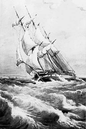
H. M. S. Rattlesnake by W. Brierly
Illustrated London News June 6, 1848
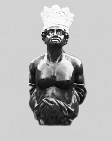
H. M.S. Rattlesnake Figurehead
Admiral Materials Laboratory,Poole, Dorset


[80] I. On the Auditory Organs in the Crustacea.
Great discrepancy prevails among the various authorities as to the true nature and position of the auditory organs in the Crustacea.
The older authors, Fabricius, Scarpa, Brandt, Treviranus, unanimously confer the title of auditory organs upon certain sacs filled with fluid which are seated in the basal joint of the second or larger pair of antennæ.
M. Milne-Edwards, in his elaborate researches upon the Crustacea,1 adheres to this determination, and describes a very elaborate tympanic apparatus in the Brachyurous genus Maia.
By the majority of the earlier writers no notice is taken of the sac existing in many genera in the bases of the first or smaller pair of antenæ. Rosenthal2 however describes this structure very carefully in Astacus fluviatilis and Astacus marinus. He considers it to be an olfactory organ, while he agrees with previous writers in considering the sac in the outer antennæ as the auditory organ.
Dr. Farre, in his admirable paper in the Philosophical Transactions for 1843, gives very good reasons for exactly reversing Rosenthal's denominations, and considering the sac in the first pair of antennæ to be the auditory organ, while the sac in the second pair is the olfactory organ. Dr. Farre doubts the existence of true auditory organs in the Brachyura.
Siebold in his Report upon the progress of the Anatomy of the [81] Invertebrata for 1843-44,3 mentions Dr. Farre's views, but seems to doubt their correctness; and they have had no better reception from Prof. Van der Hoeven4 and Erichson.5
The matter stands thus at present then. It is universally acknowledged that in the Macroura there exists in the basal joint of both the first and second pair of antennæ a sac containing a liquid, and that in the Brachyura such a sac exists at least in the second pair. According to the majority of authors the sac in the second pair is the auditory organ; and according to Rosenthal the sac in the first pair is the olfactory organ.
On the other hand, if we take Dr. Farre's interpretation, the sac in the first pair of antennæ is the auditory organ, in the second the olfactory organ.
Although the structure of the organ contained in the first pair of antennæ in the Macroura departs somewhat from the ordinary construction of an acoustic apparatus in the Invertebrata, yet the argument from structure to function, as enunciated in the paper referred to, seems almost irresistible. Still, as it has obviously not produced general conviction, I hope that the following evidence may be considered as finally conclusive.
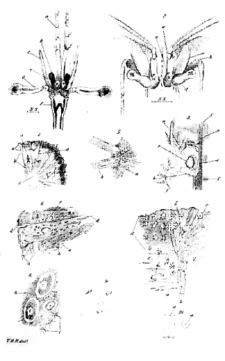
In a small transparent Crustacean (taken in the South Pacific) of the genus Palæmon (fig. 2a), the basal joint of the first pair of antennæ is thick, and provided with a partially detached ciliated spine at the outer part of its base (fig. 3a). Between this and the body of the joint there is a narrow fissure. The fissure leads into a pyriform cavity (fig. 3b), contained within a membranous sac, which lies within the substance of the joint. The anterior extremity of the sac is enveloped in a mass of pigment-granules (c): on that side of the sac which is opposite to the fissure, a series of hairs with bulbous bases are attached along a curved line (d); these are in contact with, and appear to support, a large ovoidal strongly refracting otolithe (e).
The antennal nerve (f) passes internal to, and below the sac, and gives off branches which terminate at the curved line of the bases of the hairs.
The sac is about 1/100th of an inch in length; the otolithe about 1/220th in diameter.
This structure is obviously very similar to the ordinary form of auditory apparatus in the Mollusca, &c. In Lucifer typus however we have an absolute identity.
In this singular crustacean (Pl. XIV. fig. I) the basal joint [82] of the first or internal pair of antennæ is much longer than the others, and is slightly enlarged at its base. The enlargement contains a clear vesicle (e), slightly enlarged anteriorly, but not communicating by any fissure with the exterior. It is about 1/600th of an inch in diameter. It contains a spherical strongly refracting otolithe about 1/1250th of an inch in diameter, which does not present any vibrating or rotating motion. We have here then Lucifer presenting an organ precisely similar to the auditory sacs of the Mollusca, while Palæmon offers a very interesting transition between this and the ordinary crustacean form of acoustic organ as described by Farre, and there can I think be very little doubt that the determination of the latter (as regards the Macroura at least) was perfectly correct.
Since writing the above I find that the auditory organ in Lucifer has been recognized by M. Souleyet. All that he says about it is contained in the following lines:–"Bei einigen See-krustenthieren namentlich bei der Gattung Lucifer (Thompson) habe ich ganz neuerdings an der Wurzel der innern Fühler einen kleinen runden glänzenden Körper entdeckt der mir dasselbe Organ (auditory organ) zu seyn scheint."–Froriep's Notizen, 1843, p. 83.
EXPLANATION OF PLATE XIV.
Fig. 1. The line indicates the natural size of the animal in this and the following figure: a, internal antennæ; b, external antennæ; c, basal lobe of external antennæ; d, eye; e, otolithic sac.
Fig. 2. Head of Palæmon. Letters as in fig. 1.
Fig. 3. Internal antennæ of Palæmon enlarged: a, spine; b, auditory sac; c, pigment-granules; d, curved line to which the hairs are attached; e, otolithe; f, antennal nerve.
II. On the Anatomy of the genus Tethya.
The animal which forms the subject of the present communication was found attached to rocks and stones, close to low water mark, upon the shores (skirting one of the smaller bays of Sydney harbour) of the beautiful grounds of my friend Mr. W. S. MacLeay.6
MM. Milne-Edwards and Audouin (Ann. d. Sc. Nat. 1828, tom. xv.) and Dr. Johnston (British Sponges and Lithophytes) are, so far as I am aware, the only authors who give any detailed account of the genus Tethya.
Of the two species described by the latter, T. Lyncurium approaches nearest to the present species; the only difference being that while [83] the former is yellowish white, the latter is deep red, and that the stellate bodies, scanty in the former, are very numerous in the latter.
However, pale specimens were frequent among the deep red ones–without any other apparent difference–and the presence of more or fewer stellate bodies is a mere question of degree.
MM. Edwards and Audouin describe currents traversing the "oscules" of the Tethya similar to those of a sponge. I did not observe any currents, but I do not doubt their existence.
Dr. Johnston says (op. cit p. 82), "The propagation of Tethya is by means of sporules or gemmules generated within the sarcoid matter. The latter resemble the parent sponge in miniature, but they have no distinct rind or nucleus, being composed of simple spicula woven together by albuminous matter."
I did not observe such "sporules or gemmules" in any of the specimens I examined, but, it can hardly be doubted that these bodies are merely further developments of the "ova" which I observed; and as I found spermatozoa, it will follow, that the Tethyæ are reproduced by a process of true sexual generation.
It would be most interesting to ascertain whether the "gemmules" of sponge take their origin in a similar way, and whether true spermatozoa are developed here also.
The specimens of Tethyæ observed presented several prominent tubercles upon their surface, perforated by irregular apertures, from which a liquid exuded when the animal was taken out of the water.
When there was only one or two of these tubercles, the external resemblance to some forms of Cynthia was very great.
On cutting across one of these bodies, it was seen to be solid, and composed of three distinct substances; viz. a central whitish spherical mass, a deep red cortical substance, and between these two, forming the largest part of the body, a yellowish red intermediate substance, sharply separated from both the central and cortical substances.
The two latter were united by radii of a silvery whitish colour, which ran through the intermediate yellow mass, and became lost in the cortical portion.
Small canals took their rise at the apertures already mentioned, and penetrating the cortical substance, ramified irregularly through the intermediate substance, reaching as far as, but not penetrating, the central substance. They appeared to be lined by a very delicate smooth membrane.
The general structure of the central, cortical, and intermediate portions agreed pretty closely with the description already given by Johnston.
[84] 1. The central portion.–This consists of a granular mass interpenetrated in every direction by short, cylindrical, transparent rods which form a sort of network. At the margins of the central portion, however, the rods become gathered into bundles, and they are longer and lie parallel to one another. In this form they enter the intermediate substance and form the radii before mentioned. When they reach the cortical substance, the majority of the rods diverge and become spread out; a few however remain as a bundle, and reach the edge, or even project a little beyond it.
Besides the bundles, a great number of long, solitary rods traverse the intermediate substance radially.
The rods are cylindrical, and about 1/2500th of an inch in diameter. They are all perforated by a very narrow central canal, so as to appear like minute thermometer-tubes.
2. The cortical substance consists of two zones, an inner and an outer, which pass insensibly into one another at the line of contact.
The inner is composed of a mass of thick bundles of a fibrous tissue, so interwoven that a slice presents every possible section of them. The rods penetrate this zone, and a very few of the stellate bodies are found scattered through it.
The outer zone is dense, granular, and otherwise apparently structureless. Scattered through it are great numbers of crystalline spheres beset with short conical spikes.
3. The intermediate substance.–This consists of a granular substance in which ova and stellate crystalline bodies are imbedded.
The ova are of various sizes. The largest are oval and about 1/330th of an inch in long diameter. They have a very distinct vitellary membrane, which contains an opake coarsely granular yelk. A clear circular space about 1/1600th of an inch in diameter, marking the position of the germinal vesicle, is seen in the centre of each ovum, and within this a vesicular germinal spot 1/5000th of an inch in diameter is sometimes visible, although with some difficulty, in consequence of the opacity of the yelk.
The stellate bodies are about 1/1200th of an inch in diameter: they appear to be of a similar nature to those described in the cortical substance, but they are smaller; and while the radii are proportionally long, there is hardly any centre beyond that formed by their meeting.
The granular uniting substance is composed entirely of small circular cells about 1/3300th of an inch in diameter, and of spermatozoa in every stage of development from those cells. The cell throws out a long filament which becomes the tail of the spermatozoon, and becoming longer and pointed forms, itself, the head.
[85] The perfect spermatozoa have long, pointed, somewhat triangular heads about 1/3000th of an inch in diameter, with truncated bases, from which a very long filiform tail proceeds.
It is remarkable that the ova are in no way separated from the spermatozoa, but lie imbedded in the spermatic mass like eggs packed in sand.
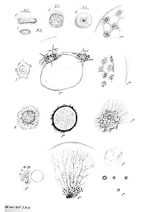
Fig. 4. Section of Tethya ; natural size: a, corticle substance; b, intermediate substance c, central substance; d, canals.
Fig. 5. Portion of central substance (a) with two of the radii (b).
Fig. 6. Segment of the cortical and intermediate substances; a, cortical substance; b, intermediate substance; c, canals cut across; d, radii.
Fig. 7. A portion of the cortical substance: a, inner fibrous portion; b, radial bundle of rods; c, stellate bodies; d, marginal homogeneous portion.
Fig. 8. A portion of the intermediate substance: a, ova; b, granular substance consisting of spermatozoa and cells; c, stellate bodies.
Fig. 9. Spermatozoa in various stages of development.
Fig.10. Longitudinal and transverse view of rods, showing the central canal,a.
Note–"Upon the Auditory Organ in Crustacea."
MM. Frey and Leuckart7 (for access to whose work I am indebted to Prof. E. Forbes since writing on this subject) express a doubt as to the correctness of any of the determinations of the auditory organ in Crustacea hitherto given. They describe a very singular organ existing in the caudal appendages of Mysis flexuosa, consisting of an oval flattened sac or cavity 1/3rd of a line in diameter, and containing an otolithe 1/4-1/6th of a line in diameter. The otolithe is discoidal, flat on the one side, umbilicated on the other, and marked with concentric lines. About two-thirds of the circumference of the otolithe are occupied by the bases of a series of glassy, stiff hairs which are inserted into the otolithe and project from it.
The otolithe is apparently composed of chitine and carbonate of lime.
No nerve was traced to this sac, but the caudal ganglion is of large size.
No similar organ exists in Palæmon, Crangon, or Squilla, but the authors compare it to the organ noticed by Souleyet in Lucifer; and notwithstanding the extraordinary position of the organ, it must be allowed that its structure goes far to support this view. It must be remembered that in some of the lower Annelida the auditory organs are situated, not in the head, but one or two rings behind it, and in Polyophthalmus every ring has its pair of eyes.–See Quatrefages Ann. d. Sc. Nat. 1850.
[86] III. Upon Thalassicolla, a new Zoophyte.
In all the seas, whether extra-tropical or tropical, through which the Rattlesnake sailed, I found floating at the surface the peculiar gelatinous bodies which are the subject of the present communication. They were the most constant of all the various products of the towing-net, which was rarely used without obtaining some of them, and which sometimes, for days, would contain hardly anything else.
The extreme simplicity of structure of these creatures was more puzzling to me than any amount of complexity would have been. The difficulty of perceiving their relations with those forms of animal life with which I was familiar, gave me rather a distaste to the study of them, and, as I now perceive, has rendered my account of their organization far less complete than I could wish it.
However, these forms seem completely to have escaped the notice of voyagers, and therefore I hope to do some service by directing the attention of future investigators to them, and by endeavouring to show what seem to me to be their relations in the scale of being.
It may not be out of place at the same time to examine what are the positive characters of those lowest classes of animal life of which this is a member.
The Thalassicolla8 is found in transparent, colourless, gelatinous masses of very various form;–elliptically-elongated, hour-glass-shaped, contracted in several places, or spherical, varying in size from an inch in length downwards; showing no evidence of contractility nor any power of locomotion, but floating passively on the surface of the water.
Now of such bodies as these there were two very distinct kinds: the one kind, consisting of all the oval or constricted, and many spherical masses, is distinguished to the naked eye by possessing many darker dots scattered about in its substance; the smaller kind, always spherical, has no dots, but presents a very dark blackish centre, the periphery being more or less clear. I will adopt the provisional name of Th. punctata for the former kind, and that of Th. nucleata for the latter, as a mere matter of convenience, and without prejudging the question as to the existence of specific distinctions.
Th. punctata. (Pl. XVI. figs. 1, 2, 3.)
The mass consists of a thick gelatinous crust containing a large cavity. The crust is structureless, but towards its inner surface minute [87] spherical, spheroidal or oval bodies are imbedded, from which the appearance of dots arises. These are held together merely by the gelatinous substance, and have no other connexion with one another. Each "spheroid" is a cell, with a thin but dense membrane, 1/200th to 1/250th of an inch in diameter, and contains a clear, fatty-looking nucleus 1/1400th to 1/800th of an inch in diameter, surrounded by a mass of granules which sometimes appeared cellæform.
This fundamental structure–a mass of cells united by jelly–like an animal Palmella, was subject to many and important varieties.
Very commonly the central part of each mass, instead of containing a single large cavity, consisted of an aggregation of clear, large, closely-appressed spaces, like the "vacuolæ" of Dujardin (figs. 2, 3, 2 a, 3 a).
Very frequently also each cell was surrounded by a zone of peculiar crystals somewhat like the stellate spicula of sponge, consisting of a short cylinder, from each end of which three or four conical spicula radiated, each of these again bearing small lateral processes (figs. 3 a, 2 b).
In another kind, much more rarely met with, the spherical cell contained a few prismatic crystals about l/1000th of an inch in length; it was of a bluish colour, and enveloped in a layer of densely packed minute granules not more than 1/15000th of an inch in diameter. Outside these there was a number of spherical bright yellow cells 1/1600th of an inch in diameter, and inclosing the whole a clear, transparent brittle shell perforated by numerous rounded apertures, so as to have a fenestrated appearance (fig. 6). There were no spicula in this kind.
In a single specimen I found a similar shell, but its apertures were prolonged into short tubules (fig. 5).
Frequently the connecting substance in which the cells were imbedded appeared to be quite structureless, but in some specimens delicate, branching, minutely granular fibrils were to be seen radiating from each cell into the connecting substance (fig. 2 b).
I have mentioned certain minute bright yellow spherical cells contained within the shell of the fenestrated kind; such coloured cells are contained in all kinds either diffused through the connecting substance or more or less concentrated round each large cell (figs. 3 a, 2 b).
Th. nucleata. (Pl. XVI. fig. 4.)
This form consists of a spherical mass of jelly as large as the middle-sized specimens of the last variety, with an irregular blackish central mass. Enveloping this and forming a zone about half the [88] diameter of the sphere there is a number of clear spaces–vacuolæ–varying in size from 1/62nd to 1/2500th of an inch, the smallest being innermost. Scattered among the vacuolæ of the innermost layer, there were many of the yellow cells, and a multitude of very small dark granules. Delicate, flattened, branching fibrils radiated from the innermost layer, passing between the vacuolæ, and in one specimen these fibrils were thickly beset with excessively minute dark granules, like elementary molecules, which were in active motion, as if circulating along the fibrils, but without any definite direction. In this case the whole body looked like a moss agate, so distinct were the radiating fibrils (4 a). Left to itself for less than an hour, however, this appearance as well as the circulation of granules vanished, and only a few scattered radiating fibrils were to be observed, the rest seeming to have broken off and become retracted.
By rolling under the compressor the outer mass could be completely separated from the central dark body, which then appeared as a spherical vesicle 1/65th of an inch in diameter (fig. 4 b), showing obscurely a granular included substance.
The membrane of the vesicle was very strong, resisting, and elastic. When burst it wrinkled up into sharp folds (fig. 4 c), and gave exit to its contents (fig. 4 d). These were–
1. A very pale delicate vesicle (nucleus?) without any contents, and measuring (but when much compressed) about 1/60th of an inch (fig. 4 d).
2. A heterogeneous mass consisting of (a) a finely granular base, (b) oil-globules of all sizes, (c) peculiar cells 1/800th to 1/1000 of an inch in diameter (4 e). Some of these had a solid greenish red nucleus about 1/3500th of an inch in diameter. Others resembled the nuclei in colour and appearance, but were larger (1/2500th of an inch), and had no cell-membrane:–were these granule cells?
Altogether the Thalassacol1a nucleata might readily be imagined to be a much enlarged condition of single cells of the Th. punctata; but I have no observations to show that it was so, nor can it be said from which of the varieties of Th. punctata the Th. nucleata arises.
The question may readily arise, Are these perfect forms? I can only say, as negative evidence, that I have never observed any trace of their further development, and that the spicula and 'shells,' and the capacity of fission, appear to afford positive grounds for believing that they are not mere transitional stages of any more highly-organized animal. If, further, it can be shown that their structure is closely allied to that of known organisms, this probability will, I think, almost amount to a certainty.
[89] What animals are there then which consist either of simple cells or of cells aggregated together, which hold the same rank among animals that the Diatomaceæ and Desmidiæ, the Protococci and Palmellæ hold among plants?
Ten years ago the general reply of zoologists would have been–none. The researches of the celebrated Berlin microscopist, Prof. Ehrenberg (wonderful monuments of intense and unremitting labour, but at least as wonderful illustrations of what zoological and physiological reasoning should not be), led to the belief that the minutest monads had an organization as complicated as that of a worm or a snail. In spite, however, of the great weight of Prof. Ehrenberg's authority, dissentient whispers very early made themselves heard, from Dujardin, Focke, Meyen, Rymer Jones, and Siebold. To these Kölliker, Stein and others–in fact, I think I may say all the later observers–have added themselves, until it really becomes a matter of duty on the part of those interested in the progress of zoology to pronounce decidedly against the statements contained in the 'Infusionsthierchen,' so far as regards anatomical or physiological facts.9
It has been shown in the first place, that a great mass of the so-called Polygastria are plants–at any rate are more nearly allied to the vegetable than to the animal kingdom. Such is the case with the Diatomaceæ and Desmidiæ, the Volvocina, the Monadina, the Vibriones, and to these we must very probably add the Astasiæa.
So utter has been the want of critical discrimination in the construction of genera and species, that Cohn, in his admirable memoir upon Protococcus pluvialis, enumerates among the twenty-one forms (to which distinct names have been given by authors) assumed by the Protococcus, no less than eight of Prof. Ehrenberg's genera. The family "Polygastria," thus cut down to less than one-half its original dimensions, contains none but animals which are either simple nucleated cells, or such cells as have undergone a certain amount of change, not sufficient however to destroy their real homology with nucleated cells.
A nucleus has been found in Euglena, Arcella, Amœba, Amphileptus, Trachelius, Bursaria, Paramæcium, Nassula, Chilodon, Oxytricha, Stylonichia, Stentor, Vorticella, Euplotes, Trichodina, Loxodes, and other genera. It may be brought out by acetic acid just like any other nucleus in Vorticella and Euglena.
[90] The animal is an unchanged cell in Euglena, in Amœba and in Opalina. In others, as the Vorticellæ, there is a more or less distinct permanent cavity in the interior of the cell which opens externally, an occurrence not without parallel among the secreting cells of insects. Certain genera, such as Nassula, have an armature of spines, but so have some of the Gregarinidæ which are unquestionably simple cells.
Contractile spaces,–cavities which appear and disappear in different parts of the Infusoria, and sometimes become filled with the ingesta,–are found no less commonly in the component cells of the tissues of many of the lower animals, and according to Cohn in the primordial cells of plants also.
The "Polygastria," then, may be justly considered to be simple cells, and to form a type perfectly comparable with Thalassicolla.
The researches of Henle, Stein, and Kölliker have made us acquainted with another form of cellular animals–the Gregarinidæ.
These are nucleated cells, without cilia, but with contractile walls, which lead an independent parasitic life in the intestines of many of the Invertebrata, principally insects.
The Gregarinidæ, like the Infusoria, are generally, if not invariably, single, solitary cells.
A third type is formed by the Foraminifera. The fate of these animals is somewhat singular. Considered to be Cephalopoda by D'Orbigny; Bryozoa by Ehrenberg; rudimentary Gasteropods by Agassiz; all careful observation tends to confirm the opinion of Dujardin, that the fabrication of their remarkable shells is essentially similar to Amœba and Arcella, both of which have been shown to be nucleated cells.
Lastly, we have the Sponges. That the tissue of the Sponges breaks up into masses, each of which is similar to an Amœba, has been pointed out by Dujardin, and confirmed by Carter and others. Dujardin, however, believing that a peculiar formless substance, "Sarcode," constitutes the tissues of the Sponges (as well as of the Infusoria and many other of the lower animals), fails to point out that they are mere aggregations of true cells.
This is not the place to discuss the important question, whether the great law developed by Schwann does or does not hold good among the whole of the lower animals. I believe that there is evidence to show that it does; that everywhere careful analysis will demonstrate the nucleated cell to be the ultimate histological element of the animal tissues; and that the "sarcode" of Dujardin, and the "formless contractile substance" of Ecker, are either cells or cell-contents, or the results of the metamorphosis of cells. Be this as it may, however, I [91] can say positively, as the result of recent careful examination, that Spongilla, Halichondria and Grantia are entirely composed of nucleated cells.
The Foraminifera and Sponges then, no less than the Infusoria and Gregarinidæ, are "unicellular" animals–animals, that is, which either consist of a single cell, or of definite aggregations of such cells, none of which possesses powers or functions different from the rest.
Using the word "unicellular" in this extended sense (as it has been used by Nägeli and others with regard to the Algæ), it may be said that there are four families of unicellular animals; in two of these, the Infusoria and Gregarinidæ, the cells are isolated; in two, the Foraminifera and Sponges, they are aggregated together.
From these considerations it appears to me that the zoological meaning and importance of the Thalassicolla punctata first become obvious. It is the connecting link between the Sponges and the Foraminifera. Allied to the former by its texture and by the peculiar spicula scattered through the substance of some of its varieties, it is equally connected with the latter by the perforated shell of other kinds. If it be supposed that a Thalassicolla becomes flattened out, and that a deposit takes place not only round the cells, but between the partitions of the central "vacuolæ," it becomes essentially an Orbitoides.10
To come to a similar understanding of the nature of the Thalassicolla nucleata, it is necessary to recur again to certain general characteristics of the reproductive processes in the unicellular animals.
If we except Tethya, a sponge,11 the ordinary reproductive elements have as yet been found in no unicellular animal.
Fission occurs in all except perhaps the Gregarinidæ. Gemmation appears to take place in the Foraminifera and Infusoria. In the Sponges the so-called ova or gemmules seem to be only a temporary locomotive condition of the cells, such as occurs in the Vorticellæ among the Infusoria, and the Protococci among plants.
But in all (except the Foraminifera) a process of multiplication by [92] endogenous development occurs, and would seem in some cases to represent sexual propagation. Now the mode of this endogenous multiplication presents remarkable features of similarity in the Infusoria, the Gregarinidæ, and the Sponges.
There is a certain period in the existence of Vaginicola crystallina, when, gorged with food stored up in the shape of fat granules, &c., within the cavity of its cell-body, it becomes sluggish and eventually still. The body contracts and becomes rounded, and the transparent case closes in and seals up its inhabitant. Eventually long processes are developed from the body, and it takes on the form of the genus Acineta of Ehrenberg. After a while a new life stirs within this chrysalis-like form, and the contained mass gives rise successively (by a sort of fission) to young ciliated bodies, which leave the Acineta and become Vaginicolæ.
In a similar manner Vorticella microstoma becomes Podophrya fixa; but sometimes the changed Vorticella has no stalk, and then is the Actinophrys of some authors (not A. Sol). It is not known in what way the embryos are brought forth here, but it is a very significant fact that both the stalked and unstalked forms have been observed to conjugate.
Epistylis presents similar phænomena.
The Actinophrys Sol, to which more particular reference will be made by and by, has been observed to conjugate, but it is not absolutely known to arise by the metamorphosis of any Vorticella, though there is every probability in favour of the supposition that it does.
The Gregarinidæ pass through similar changes. Two forms of these creatures are known; the one consisting of protean nucleated cells, the other of motionless spherical sacs, containing a vast number of minute bodies resembling Naviculæ in shape, and thence called "Navicella-sacs." Now, according to Stein, although the fact has been doubted by others, the "Navicella-sacs" result from the conjugation of two Gregarinæ, which have become motionless and filled with an accumulation of granules.
Certain it is that the Navicellæ are developed within the granular mass like embryo-cells within the yelk, and that when freed by the bursting of the Navicella-sac they become Gregarinæ.
Lastly, in the freshwater sponge (Spongilla), which consists of an aggregation of nucleated protean cells like a mass of Gregarinæ, a certain number of the cells at various points scattered through the substance of the Spongilla become motionless and distended with granules, and receiving first a membranous and then a siliceous investment, constitute the "seed-like bodies."
[93] From Mr. Carter's account it would appear that when the "seedlike body" germinates its cells burst, and their granular contents become mixed. Subsequently protean cells, like the ordinary sponge-cells, make their appearance pari passu with the disappearance of the granules.
Supposing this account to be correct, the conjugation in Spongilla would be perfectly analogous to that of the Desmidiæ and Diatomaceæ, while in the Infusoria and Gregarinidæ it would resemble that of Zygnema.
Generalizing the above details (full authority for which may be found in the appended list of works), we may say that with the exception of the Foraminifera, about whose reproductive processes nothing is as yet known, the Protozoa all reproduce their kind by a process of endogenous development which is accompanied by greater or less changes in the structure and powers of the reproducing cell. We may add that in many cases these changed cells have been observed to conjugate, previous to the occurrence of the endogenous development.
Bearing all these facts in mind, let us return to Thalassicolla nucleata. If the Th. punctata answer to a mass of sponge-cells, or an aggregation of Gregarinæ, is it not possible that the Th. nucleata may answer to the altered reproductive cell? I have shown that the Th. nucleata may very possibly be nothing more than a separated and enlarged cell of Th. punctata, and this possibility upon structural grounds becomes, I think, converted into probability, if Th. nucleata be compared with Actinophrys Sol, which there is every reason to believe is the reproductive stage of one of the Vorticellinæ.
Actinophrys Sol is a spherical gelatinous mass consisting of an internal dark granular portion and a clearer external zone from which many radiating threads are given off. Vacuolæ are scattered through the substance, larger in the external zone, smaller and more irregular in the interior.
If the animal is much compressed, nuclei and nucleated cells are forced out from its interior.
Finally, two specimens of Actinophrys have been observed to fuse together and become one.
It is unnecessary to point out the perfect analogy between Actinophrys and Thalassicolla nucleata, with one exception, that the large internal cell was not observed in Actinophrys–a circumstance which might readily occur if it were delicate, even though it existed.
The argument derived from this analogy becomes still more strengthened if we turn to the excellent account of Noctiluca–a marine phosphorescent body which has long been a zoological puzzle [94]–by M. de Quatrefages. For the details I must refer to that observer's paper in the 'Annales des Sciences,' but I may state that its structure is essentially similar to that of Thalassicolla nucleata, supposing that the latter had given exit to its central cell by a depression at one point of its surface. Noctiluca, however, appears to feed after the manner of Actinophrys, and perhaps conjugates also, as M. de Quatrefages "has met with double individuals two or three times." This he considers an evidence of spontaneous fission; but further observation might have reversed this judgement, as it did that of Kölliker with regard to Actinophrys.
From the invariable adhesion of grains of sand to one part of the surface of Noctiluca, it would seem to be set free from some unknown fixed form which is probably analogous in its structure to Thalassicolla punctata.
To sum up the different lines of argument it may be said–
1. That the Thalassicolla punctata is not an exceptional form of animal life, but belongs to the same great division as the Sponges, Foraminifera, Infusoria, and Gregarinidæ,–the Protozoa or unicellular animals.
2. That the Protozoa have definite characters as a class, which are–
a. That they are either simple nucleated cells or aggregations of such cells, which are not subordinated to a common life.
b. That they have a mode of reproduction consisting in an endogenous development of cells, preceded by a process analogous to the conjugation of the lower plants.
3. That the Thalassicolla nucleata closely resembles Actinophrys Sol, which is known to conjugate, and which there is great reason to believe is the reproductive stage of one of the Vorticellinæ.
4. That as Th. punctata is one of the Protozoa, it most probably has a reproductive stage.
5. That Th. nucleata might readily be derived from such an alteration in one of the cells of Th. punctata as occurs in the sponge-cells when they go to form the seed-like body, or in the Gregarina-cells when they become "Navicella-sacs."
6. That Thalassicolla nucleata is essentially similar in structure to Noctiluca.
Finally, I may be permitted to say, that no one can be more fully conscious than myself of the slender and hypothetical grounds on which some of these conclusions rest. My chief purpose has been [95] merely to show the tendency of the evidence now extant as clearly and broadly as possible;–rather to draw out a brief than to pronounce a judgment.
The following are the authorities referred to in the text:–
Von Siebold. Vergleichende Anatomie d. Wirbellosen Thiere, p. 7-25.
––Ueber einzellige Pflanzen u. Thiere. Siebold und Kölliker's Zeitschrift, 1849.
Dujardin. Histoire naturelle des Zoophytes Infusoires.
Cohn. Nachträge zur Naturgeschichte des Protococcus pluvialis, Nova Acta Acad. Nat. Cur. 1850.
Pineau. Sur le Développement des Infusoires. Annales des Sciences, 1845.
Stein. Untersuchungen über die Entwickelung d. Infusorien. Wiegmann's Archiv. 1849.
––. Ueber die Natur d. Gregarinen. Müller's Archiv, 1848.
Kölliker. Ueber die Gattung Gregarina. Siebold und Kölliker's Zeitschrift, 1848.
––. Das Sonnenthierchen. Ditto, 1849.
Quatrefages. Observations sur les Noctiluques. Annales des Sciences, 1851.
Hogg. Upon Spongilla. Trans. Linn. Soc. vol. xviii.
Carter. On Spongilla. Annals of Nat. Hist. 1849.
l Hist. Nat. des Crustacés. Suites à Buffon. 2 Ueber die Geruchsorganen d. Insekten. Reil's Archiv, B. x. 1811. 3 Müller's Archiv, 1845. 4 Handbuch d. Zoologie, p. 597. 5 Erichson's Archiv, 1844. 6 It is not necessary for me to speak of Mr. MacLeay's singular acquirements and acumen; but I cannot refrain from taking this opportunity of expressing my deep sense of the benefit I have derived from his advice and assistance–always most readily offered. 7 Beiträge zur Kenntniss Wirbelloser Thiere. 8 [Thalassa], the sea; [kolla], jelly, glue. 9 That the above assertions will be considered by the majority of English readers to be unwarrantably severe, and considering the relative standing of the Professor and his critic, possibly impertinent, is no more than is to be expected. I can only beg to disclaim all mere iconoclastic tendencies, and refer to a comparison of Prof. Ehrenberg's works with facts for my justification. 10 Dr. Carpenter, to whom I communicated these observations, writes to me: "As far as I can understand them, the bodies described (if perfect non-embryonic forms) seem to constitute that kind of connecting link between Sponges and Foraminifera, which the relative position I have assigned to them would lead me to expect. It is interesting to remark that the cullender-like skeleton of certain Foraminifera is extremely like in its appearance to a fragment of the shell of an Echinus, or to the plates contained in the integument of a Holothuria, and we know that these begin with a network of spicules. Consequently there is not by any means so great a distinction between the spicular skeleton of a sponge and the cullender-like skeleton of an Orbitolina as might at first sight appear." 11 See Annals of Nat. History, S. 2, vol. vii. p. 370.
|
THE
HUXLEY
FILE
|
| ||||||||||||||||||||||||||||||||||||||||||||||||||||||